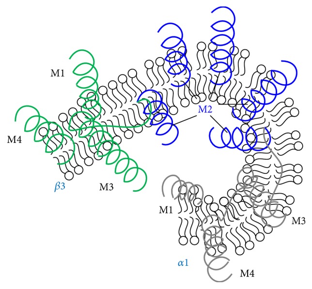
Figure 3.

GABAAR modulation patterns of transmembrane domain, a homology model of the transmembrane domains of a GABAAR showing the five-M2-helix domains forming the chloride ion channel (blue) and M1, M3, and M4 helices for single α1 (grey) or β3 (green) subunit. The helices may embed into the postsynaptic membrane in mammalian CNS.
