
Figure 2. EA.hy926 cell monolayer was intact after infection with DENV2.
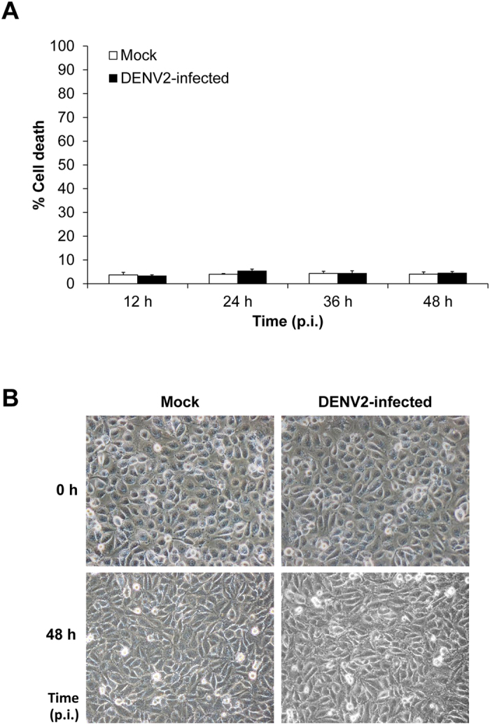
(A) Cell death was quantitated by flow cytometry using Annexin V/propidium iodide co-staining. Percentage of cell death was calculated using the formula: % cell death = [(no. of total cell death (apoptosis + necrosis)/no. of total cells) × 100%] (n = 3 independent experiments). (B) At 0 to 48 h post-infection, the morphology of mock-control and DENV2-infected cells was analyzed using an inverted light microscope. (Original magnification was 400X).