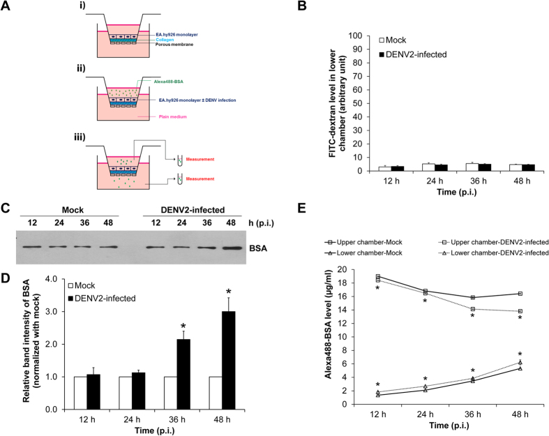
Figure 3. Integrity of EA.hy926 cell monolayer was intact, whereas albumin transcytosis was increased in DENV2-infected cells.
(A) A schematic diagram of the Transwell-based transcytosis assay. i) EA.hy926 cells form a monolayer on a collagen-coated membrane filter. ii) Alexa488-BSA was added into the insert well (upper chamber) of the Transwell chamber. iii) Alexa488-BSA remained in the upper chamber and that passed through endothelial monolayer into the lower chamber were measured. (B) The integrity of endothelial cell monolayer was evaluated by in vitro vascular permeability assay. Level of the permeated dextran conjugated with FITC (FITC-dextran) in the lower chamber, which reflects degree of permeability of the Transwell, was measured by SynergyHT microplate reader (C,D) BSA passed through the cells into the lower chamber was analyzed by Western blotting using rabbit polyclonal anti-BSA as the primary antibody. (E) Alexa488-BSA remained in the upper chamber and that passed through endothelial monolayer into the lower chamber were measured by fluorometer. (n = 3 independent experiments both Western blot analysis and fluorometry; *p < 0.05 vs. mock-control).

