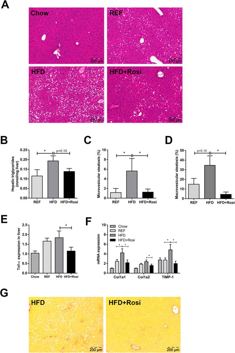
Figure 3. Effects of rosiglitazone intervention on NAFLD development.
(A) Representative photomicrographs of HE-stained liver sections of REF, HFD and HFD + Rosi. (B) Biochemical analysis of hepatic triglyceride content. Histological quantification of (C) microvesicular steatosis and (D) macrovesicular steatosis show that steatosis was ameliorated by rosiglitazone compared with HFD (n = 7–10/group). (E) TNFα gene expression in liver was diminished in rosiglitazone-treated mice (n = 7–8/group). (F) Gene expression of fibrotic genes determined by RT-PCR. Rosiglitazone reduced HFD-induced expression of Col1a1, Col1a2 and TIMP-1. (G) Onset of fibrosis in Sirius Red-stained liver cross-sections in HFD mice, but not in HFD + Rosi. Pictures are shown in magnification x100. Data are mean ± SEM, *p < 0.05. Mean expression of RT-PCR data was set 1 for chow-fed mice.

