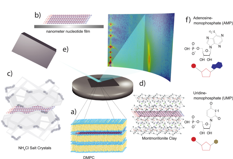
Figure 1. Schematics of systems studied, the scattering geometry, and AMP and UMP molecules.
Four environments were investigated: (a) nucleotides confined between lipid bilayers made of DMPC, (b) thin films of nucleotides applied on silicon wafers, (c) nucleotides in NH4Cl, and (d) nucleotides confined in Montmorillonite clay. (e) Two-dimensional X-ray diffraction maps were recorded to capture signals of the pure materials and signatures of the organization of the nucleotides. (f) Structures of the adenosine monophosphate (AMP) and uridine monophosphate (UMP) molecules.

