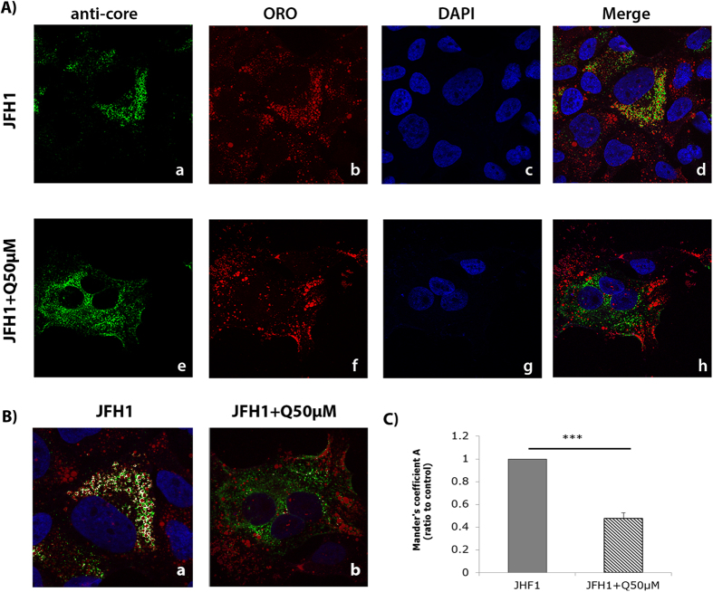
Figure 5. Effect of quercetin on the HCV core protein subcellular localization.
(A) Huh-7.5 cells were infected with JFH1 and further treated with either the vehicle (DMSO), (a) or with quercetin for 48 h (e). Core protein was detected using a specific antibody (a–e), LDs were stained with ORO (b–f) and nucleus with DAPI (c-g). Overlay images are shown in d and h. Images were taken using a confocal microscope (LSM700 Meta, Zeiss) equipped with a 63x objective. (B) Colocalization pictures. Core protein and LDs colocalization were analyzed using the Imaris software 3D Colocalization. The presence of white pixels represents the green pixels colocalized with the red pixels. (C) Colocalization statistical analysis obtained by Imaris software was analyzed using the Mander’s coefficient value. The results are means ± SD obtained from three independent experiments (25 cells were analyzed) (***p < 0.001).

