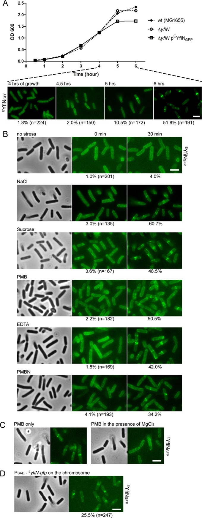
FIG 3 .

YfiN relocates to the midcell in response to envelope stress in E. coli. (A) Growth curves of E. coli wild-type (strain MG1655) cells, ΔyfiN cells, or ΔyfiN cells expressing EYfiNGFP from pBAD30 with the inducer arabinose added at time zero. The localization of EYfiNGFP at selected time points is shown in the images below. (B) EYfiN relocation to the midcell upon envelope stress. E. coli ΔyfiN cells expressing EYfiNGFP were exposed to the indicated stresses. The same microscope fields were photographed before and 30 min after exposure to no stress (LB medium only), osmotic upshift (LB with 250 mM additional NaCl or 10% sucrose), or OM permeabilization (LB with 2.5 µg/ml PMB, 10 mM EDTA, or 200 µg/ml PMBN). For each stressor, the same field of cells was observed to count the fraction of cells showing midcell foci. (C) Effect of added Mg2+ on PMB-triggered EYfiN relocation. E. coli ΔyfiN cells expressing EYfiNGFP were exposed to 2.5 µg/ml PMB for 30 min in the absence and presence of 10 mM MgCl2. (D) Localization of EYfiNGFP expressed from the chromosomal inducible promoter. An E. coli yfiR::kan mutant strain, in which EyfiN-gfp is carried under the PBAD promoter on the chromosome, was grown with arabinose at 30°C for 4 h and exposed to 250 mM NaCl for 30 min.
