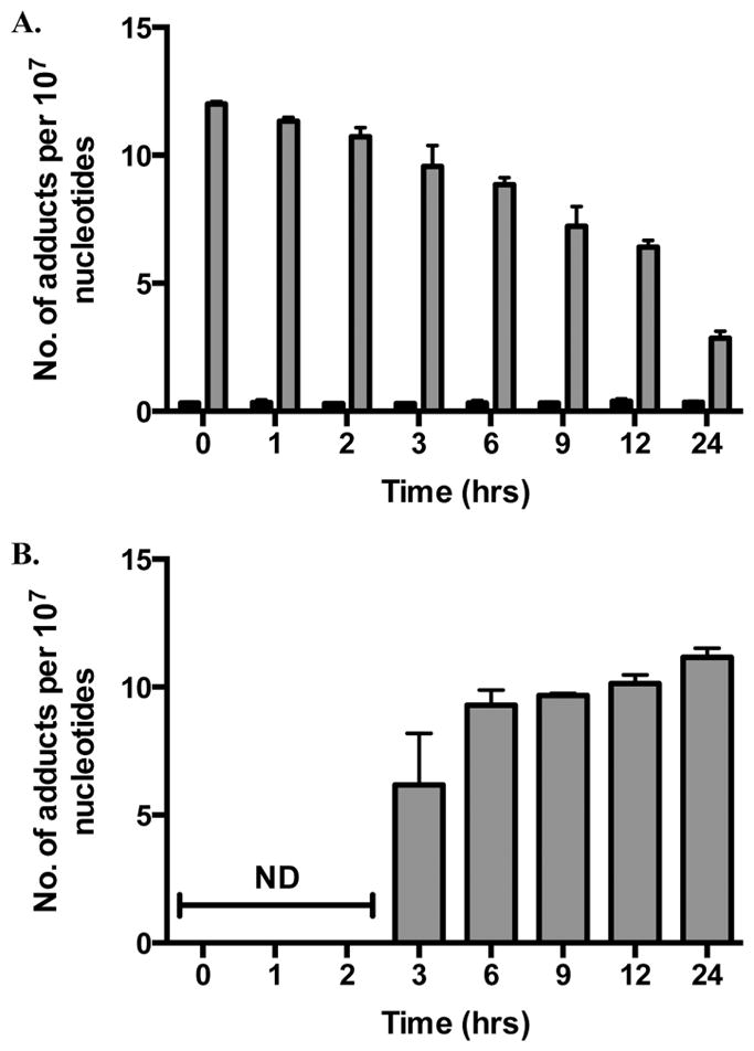
Figure 7.

M1dG and 6-oxo-M1dG levels in synchronized HEK293 cells after treatment with adenine propenal for 1 h followed by incubation for the indicated times. Data are shown for vehicle-treated (black) and adenine propenal-treated (gray) cells and indicate levels of M1dG (A) and 6-oxo-M1dG (B) in the nucleus. Data represent the mean ± SD of triplicate determinations. The data shows that during the first 3 h following adenine propenal treatment M1dG levels declined by approximately 20%, whereas a 73% decline was observed between the 3 and 24 h time points. An almost equivalent (80%) increase in the levels of 6-oxo-M1dG was seen between the 3 and 24 h time points.
