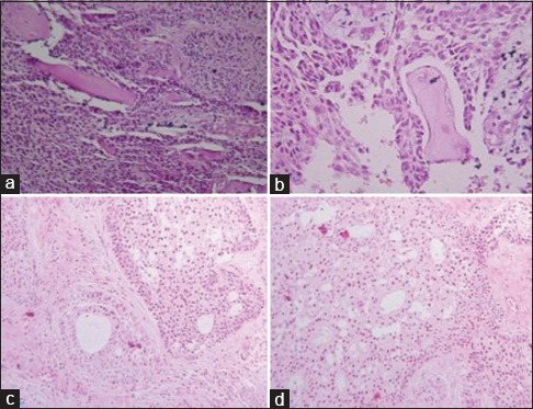
Figure 3.

(a and b) Matrix material consistent with dentinoid. (c and d) Zone of follicular pattern of ameloblastoma demonstrating peripheral ameloblastic cells and duct-like pattern. The central zone exhibiting granular cells and foci of cystic degeneration.
