Figure 5.
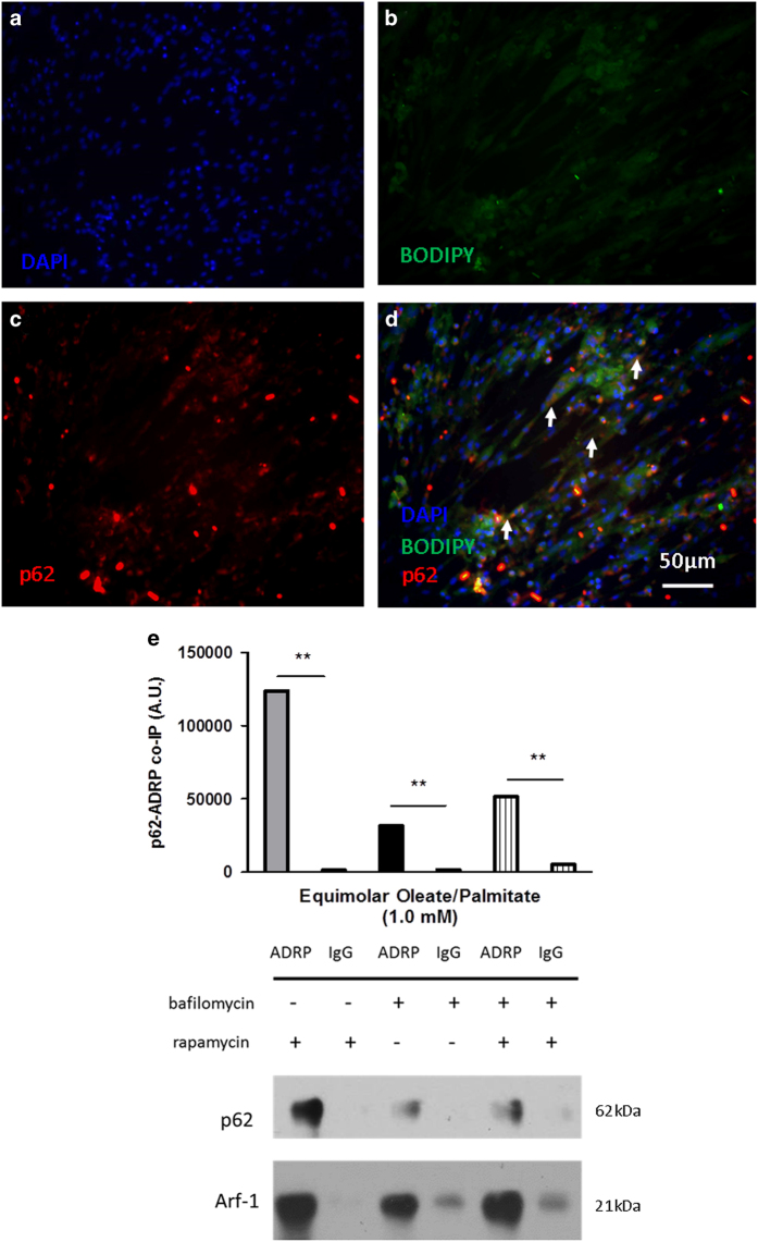
p62 associates with lipid droplets with the combination treatment of both rapamycin and bafilomycin. Immunofluorescence of cells stained with (b) BODIPY 493/503 (neutral lipid stain) and (c) TRITC (p62). (a) DAPI was used for nuclear staining. Cells were treated with both rapamycin and bafilomycin. White arrowheads in panel (d) indicate areas of colocalization between p62 and lipid droplets. All images were taken at ×40 magnification. (e) Immunoblotting for co-immunoprecipitation between p62 and ADRP in the presence of rapamycin, bafilomycin, or both. IgG groups represent negative controls; immunoblotting for Arf-1, known co-immunoprecipitate of ADRP, represent the positive controls. All cells were treated with 1.0 mM oleate/palmitate (**P<0.01).

