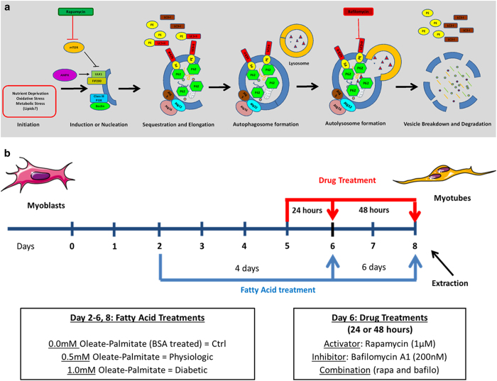
Figure 8.
Modulation of autophagy and experimental design. (a) Autophagy is triggered by various stresses, including, but not limited to, nutrient deprivation, oxidative stress, and metabolic stress. To assess autophagic flux, we measured levels of two proteins: LC3, microtubule-associated protein light chain 3, and p62/SQSTM1 (Sequestosome1 complex), an adapter protein which acts as a substrate for the autophagy pathway. Two drugs, rapamycin and bafilomycin A1, were utilized to activate and inhibit the autophagy pathway, respectively. The autophagy pathway proceeds through six major steps: (1) Initiation, (2) Induction, (3) Elongation, (4) Autophagosome formation, (5) Autolysosome formation, and (6) Vesicle breakdown and degradation. (b) Summary of experimental design. Cells were treated with various fatty acid concentrations (0.0 mM oleate/palmitate=Control; 0.5 mM oleate/palmitate=Physiologic; 1.0 mM oleate/palmitate=Diabetic) and either rapamycin (1 uM), bafilomycinA1 (200 nM) drugs, or a combination of both.

