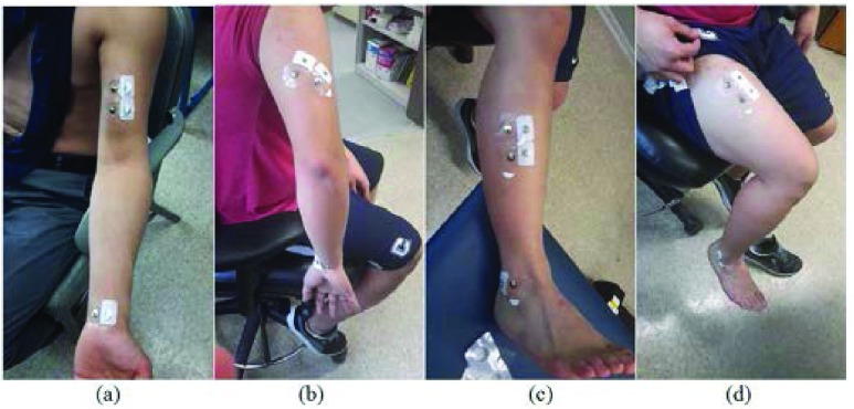
FIGURE 2.
Sample images of where the electrodes were placed for each muscle-contraction test. Note that the electrodes presented in these images were not the ones that were used for the actual tests. (a) Biceps brachii; (b) Triceps brachii (long head); (c) Tibialis anterior; (d) Quadriceps femoris (rectus femoris).

