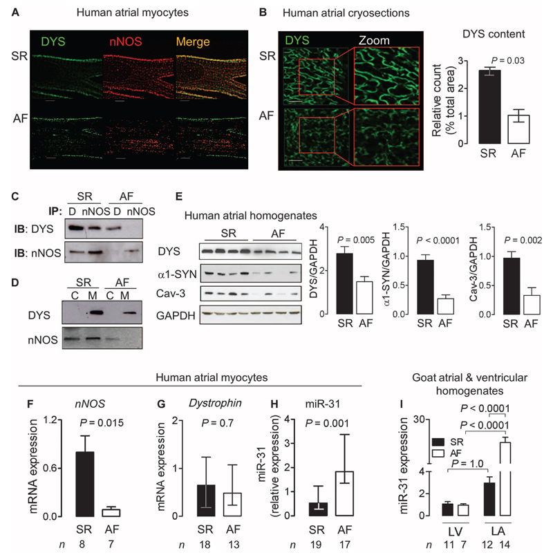
Fig. 3. Loss of dystrophin and miR-31 up-regulation in human AF.
(A and B) Immunostaining for nNOS and dystrophin (DYS) in atrial myocytes or cryosections from patients in SR or AF. Scale bars, 20 μm. Data in the bar graph are medians with interquartile ranges (n = 4 per group). P value was determined by Mann-Whitney U test. (C) Dystrophin (DYS or D) and nNOS immunoprecipitation (IP) and immunoblotting (IB) (n = 5 biological repeats per group). (D) Immunoblots of dystrophin and nNOS in the membrane (M) and cytosolic (C) fraction of atrial tissue homogenates from patients in SR or AF (n = 4 biological repeats per group). (E) Immunoblots for dystrophin, α1-syntrophin (α1-SYN), and caveolin-3 (Cav-3) in atrial tissue from patients in SR (n = 9) or AF (n = 10). Data are averages ± SEM. P values were determined by unpaired t test. (F) nNOS mRNA expression in atrial myocytes from patients in SR or AF. (G) Dystrophin mRNA expression in atrial myocytes from patients in SR or AF. (H) miR-31 expression in atrial myocytes from patients in SR or AF. Transcripts were normalized to GAPDH (dystrophin and nNOS) or snoU6 (miR-31). Data are averages ± SEM (F) or medians and interquartile ranges (G and H). P values were determined by unpaired t test (F) or Mann-Whitney U test (G and H). (I) miR-31 expression in left ventricular (LV) and left atrial (LA) tissue from goats in SR (n = 11) or AF (n = 7). Data are averages ± SEM. P values were determined by one-way ANOVA with Bonferroni correction.

