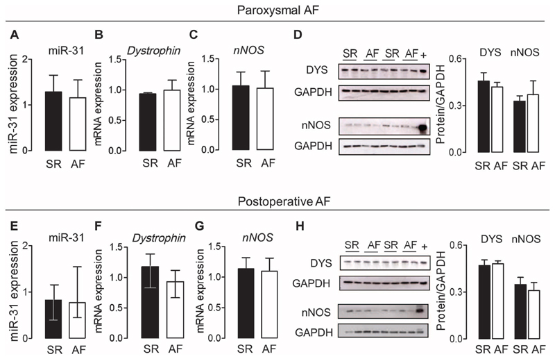
Fig. 4. Atrial miR-31, dystrophin, and nNOS in patients with paroxysmal AF and in those who developed AF after cardiac surgery.
(A to D) Expression of miR-31 (A), dystrophin (B), and nNOS (C) mRNA and dystrophin and nNOS immunoblots (D) in right atrial tissue from patients with paroxysmal AF (n = 5 to 8) or SR (n = 6 to 8). The positive control for nNOS and dystrophin (indicated by +) is murine skeletal muscle. Data are averages ± SEM. (E to H) Expression of miR-31 (E), dystrophin (F) and nNOS (G) mRNA and dystrophin and nNOS immunoblots (H) in right atrial tissue from patients in SR who developed AF after cardiac surgery (n = 18 to 12) versus those who remained in SR postoperatively (n = 8 to 11). The positive control for nNOS and dystrophin (indicated by +) is murine skeletal muscle. Data are medians and interquartile ranges (E and F) or averages ± SEM (G and H). All comparisons (A to H) between SR and AF were not significant [unpaired t test or Mann-Whitney U test (E and F)].

