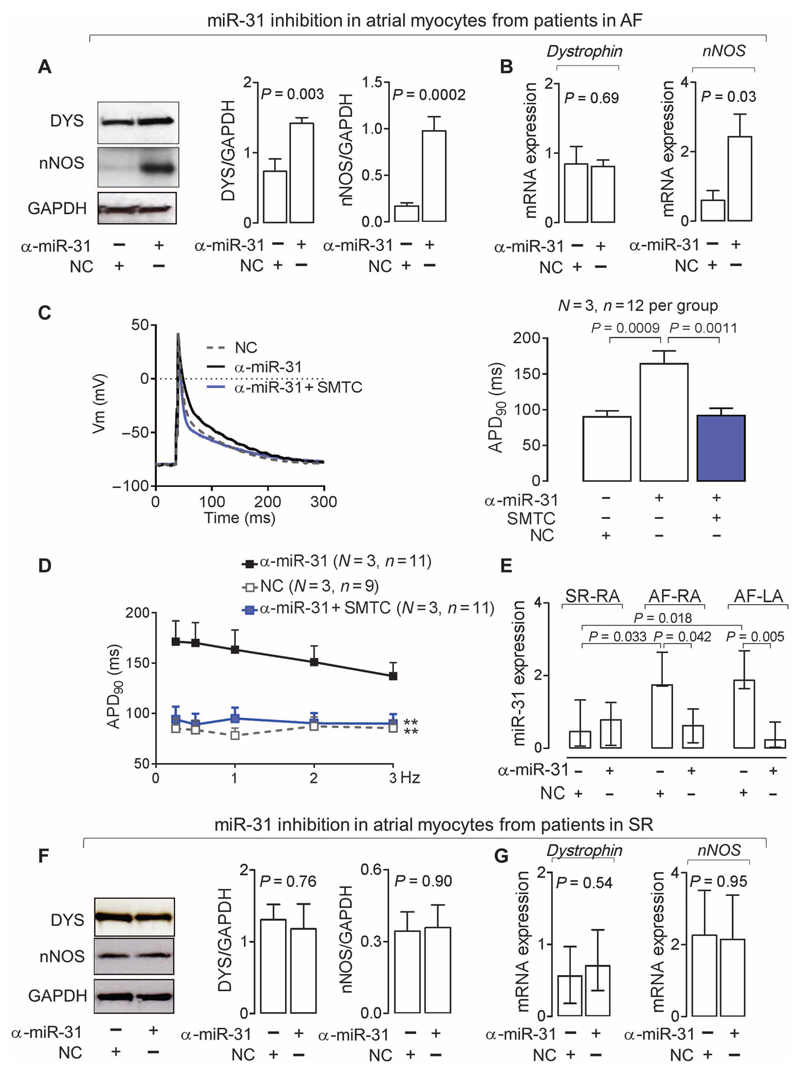
Fig. 6. Inhibition of miR-31 recovers nNOS and dystrophin and restores APD in AF.
(A and B) Effect of an miR-31 inhibitor (α-miR-31; n = 9) or a nontargeting negative control (NC; n = 5 to 9) on nNOS or dystrophin protein and mRNA in atrial myocytes from patients with AF. Data are averages ± SEM. P values were determined by paired t test (A) or unpaired t test (B) when the tissue sample was not sufficient to provide aliquots for paired comparisons of miR-31 inhibition and NC. (C) APD90 of atrial myocytes from patients with AF treated with α-miR-31 in the presence or absence of the nNOS inhibitor SMTC. Left: representative data traces. Right: Averages ± SEM. P values were determined by one-way ANOVA with Bonferroni correction. (D) APD90 rate-dependent adaptation in atrial myocytes from patients with AF treated with α-miR-31 in the presence or absence of SMTC. Data are averages ± SEM. **P < 0.01 versus α-miR-31; P < 0.0001 for the interaction between treatment and frequency (not shown), by two-way repeated-measures ANOVA with Bonferroni correction. (E) Effect of α-miR-31 or NC on miR-31 expression in atrial myocytes from patients with AF (n = 4 to 6) or SR (n = 5 to 6). Data are medians and interquartile ranges. P values were determined by Kruskal-Wallis with Dunn’s correction. (F and G) Effect of α-miR-31 or NC on protein (F) or mRNA expression (G) of dystrophin (protein, n = 7 per group; mRNA, n = 6 per group) or nNOS (protein, n = 5 to 6; mRNA, n = 4 to 5 per group) in atrial myocytes from patients in SR. Data are averages ± SEM except for dystrophin in (G), which are medians and interquartile ranges. P values were determined by unpaired t test or Mann-Whitney U test [for dystrophin in (G)].

