Figure 7.

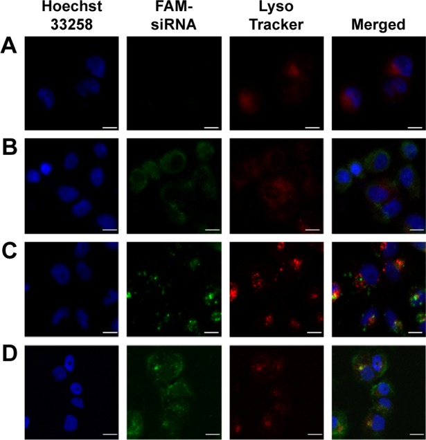
CLSM images of the intracellular distribution of FAM-siRNA.
Notes: For each panel, images from left to right show nuclei stained by Hoechst 33258 (blue), FAM-siRNA (green), lysosome stained with LysoTracker (red) and merged images. MCF-7/ADR cells were incubated with naked siRNA (A), LR (B), PSLR (C), and EPSLR (D) for 4 hours. Scale bar =10 μm.
Abbreviations: CLSM, confocal laser scanning microscopy; LR, liposome–siRNA complexes; siRNA, small interfering RNA; PSLR, PEGylated LR; EPSLR, PSLR-conjugated anti-EphA10 antibody.
