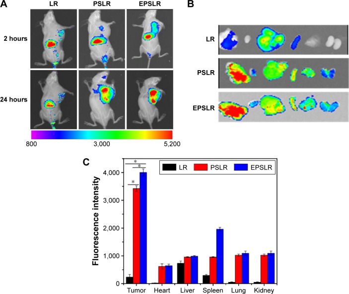
Figure 9.
In vivo fluorescence images of nude mice.
Notes: (A) Nude mice bearing MCF-7/ADR cells after tail vein administration of DIR-loaded LR, PSLR and EPSLR. (B) Ex vivo fluorescence images of tumors and organs collected at 24 hours postinjection of LR, PSLR, and EPSLR. (C) Quantification of excised organs and tumor uptake characteristics of nanocomplexes. Uptake expressed as fluorescence per mm2 of tumor and organs. Data expressed as mean values ± SD (n=3, *P<0.05).
Abbreviations: LR, liposome–siRNA complexes; siRNA, small interfering RNA; PSLR, PEGylated LR; EPSLR, PSLR-conjugated anti-EphA10 antibody; SD, standard deviation; DIR, 1,1′-dioctadecyl-3,3,3′,3′-tetramethylindotricarbocyanine iodide.

