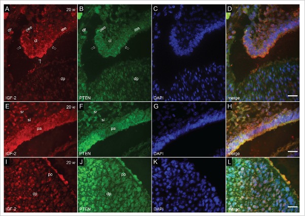
FIGURE 3.
Co-localization of IGF-2 and PTEN by double immunofluorescence in human incisor tooth germ in the 20th week of development (late bell stage). (a-d) Detail of cervical loop (a) Moderate expression of IGF-2 in the cervical loop and adjacent parts of enamel epithelia; (b) Mild expression of PTEN in the cervical loop; (c) DAPI staining of nuclei; (d) Merging of a+b+c (Magnification: ×100; scale bar: 10 µm). (e-h) Detail of enamel organ at the site of future cusp tip (e) Moderate to strong expression of IGF-2 in pre-ameloblasts. Moderate expression of IGF-2 in stratum intermedium; (f) Moderate to strong expression of PTEN in pre-ameloblasts. Mild expression of PTEN in stratum intermedium; (g) DAPI staining of nuclei; (h) Merging of e+f+g (Magnification: ×100; scale bar: 10 µm). (i-l) Detail of dental papilla at the site of future cusp (i) Mild expression of IGF-2 in pre-odontoblasts; (j) Moderate expression of PTEN in pre-odontoblasts; (k) DAPI staining of nuclei; (l) Merging of i+j+k (Magnification: ×100; scale bar: 10 µm).

