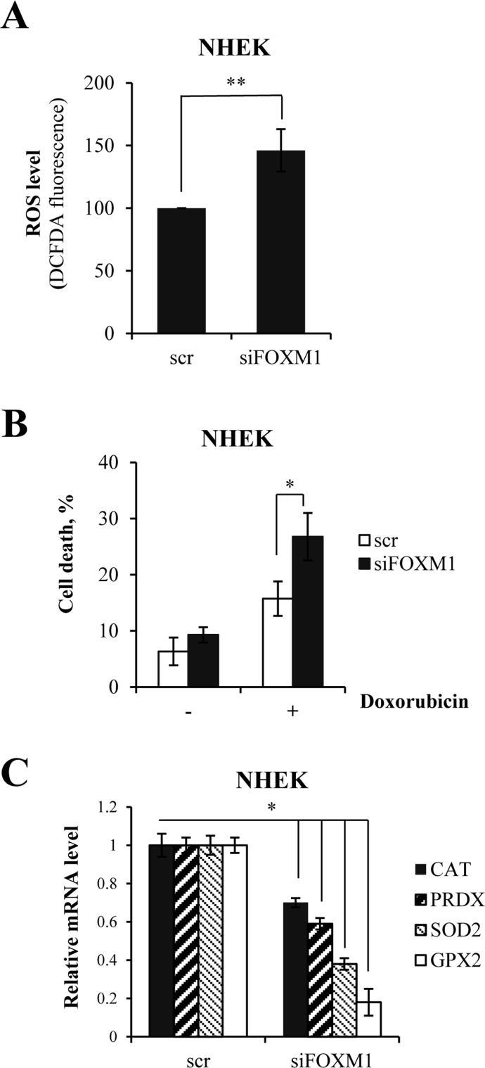
Figure 3. FOXM1 regulates oxidative stress and ROS-mediated cell death in keratinocytes.
(A) Cells were silenced for FOXM1 for 96 h. ROS levels were then measured by FACS. Values reported are the average ± SD of three independent experiments. **p-value <0.01 by Student's t-test. (B) Cells were silenced for FOXM1 for 96 h and treated with 1 μM doxorubicin. The percentage of sub-G1 events was measured by FACS at 24 h after treatment. Values reported are the average ± SD of three independent experiments. *p-value <0.05 by Student's t-test. (C) Cells were silenced for FOXM1 for 96 h, after which the relative expression levels of CAT, PRDX, SOD2, and GPX2 were determined by qRT-PCR. Values reported are the average ± SD of two independent experiments. *p-value <0.05 by Student's t-test.

