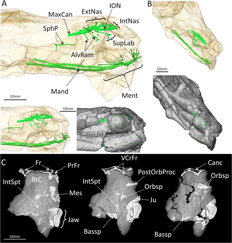
Fig 3. Three-dimensional reconstruction of the maxillary canal, mandibular canal, sphenopalatine canal and brain-case of Choerosaurus dejageri.
A, Lateral view with the skull transparent (top) and non-transparent (below). B, oblique view with the skull transparent (top) and non-transparent (below). The trigeminal nerve occupies the majority of these canals [52, 53], and the identification of trigeminal branches is based on the name of the corresponding nerve in mammals. C, CT images of the transverse section throught the braincase of Choerosaurus dejageri (from left to right: rostral to caudal sections). Abbreviations: AlvRam, alveolar rami; Bassp, basiosphenoid; BrC, braincase; Canc, cancellous bone; ExtNas, external nasal rami of the infraorbital nerve; Fr, frontal; IntNas, internal nasal rami of the infraorbital nerve; IntSpt: interorbital septum; ION, infraorbital nerve; Jaw, lower jaw bones; Ju, jugal; Mand, mandibular rami; Mes, mesethmoid; Ment, mental foramina; MxCan, maxillary canal; Orbsp, loose orbitosphenoid; PrFr, prefrontal; PostOrbProc, loose postorbital process; SphP, sphenopalatine rami; SupLab, supralabial ramus of the infraorbital nerve; VCrFr, ventral crests of the frontal. Scale bar: 10 mm.

