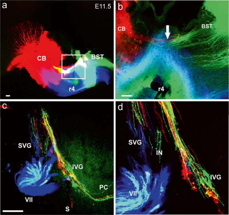
Fig. 1.

Early growth of afferents and efferents to the ear. Top images (a, b) show the injection sites of three different lipophilic dyes into the hemisected brainstem of an 11.5-day-old mouse embryo. Green is NeuroVue Jade, red is NeuroVue Orange, blue is NeuroVue Maroon. Square in (a) indicates position of (b). Note how brainstem (BST) and cerebellar (CB) afferents enter the VIII nerve (arrow in b). The whole-mounted ear (c, d) shows afferents filled from the cerebellum and brainstem (red and green) as well as efferents (blue to extend toward the targets). The higher power image shows the segregation different vestibular ganglion neurons. IVG inferior vestibular ganglion, PC posterior canal crista, S saccule, SVG superior vestibular ganglion, VII facial nerve, r4 rhombomere 4. Bar indicates 100 μm. Modified after [38]
