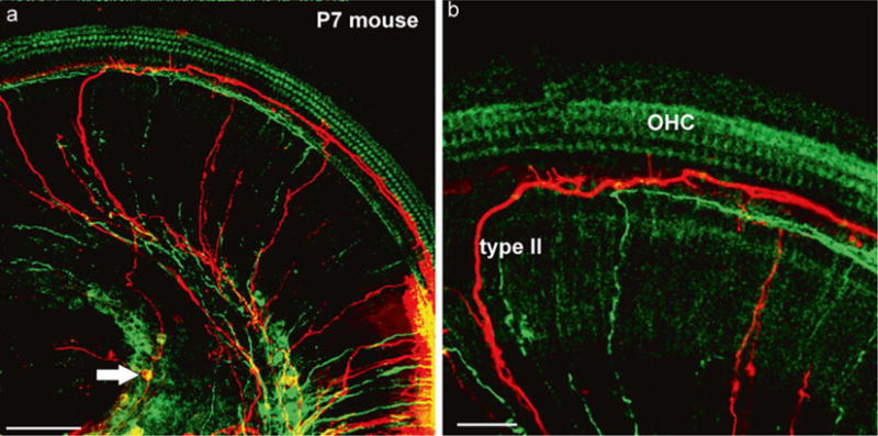
Fig. 3.

Dye labeling of afferents and efferents using peripheral applications. Two different lipophilic dyes (green, red) (a) were inserted next to each other in the dissected apex of a 7-day-old mouse. Note that both green and red (arrow) spiral ganglion cells are labeled next to the injection site (lower right in a). Two type II afferents are filled toward the apex before the turn into radial fibers and reach the spiral ganglion neurons (arrow in a). (b) The higher power image shows the fine branches emanating from type II fibers to reach the area of inner hair cells. Bar equals 100 μm in (a), 50 μm in (b). Modified after [65]
