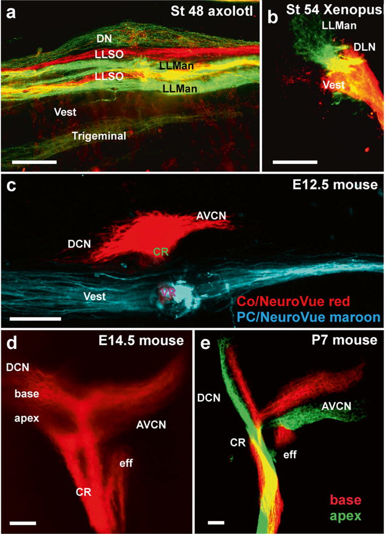
Fig. 4.

Afferent projections revealed by dextran amine and lipophilic dye tracing. (a, b) Dextran amine can be used effectively to trace different sensory organ innervation to their CNS target. The axolotl brainstem shows the termination of mandibular (LLMan) and supraorbital (LLSO) afferents in two distinct fascicles that may represent the different hair cell polarity. In contrast, afferents from the electroreceptive ampullary organs terminate overlappingly in the dorsal nucleus (DN). Frogs do not have electroreception and their lateral line projections are segregated from vestibular (Ves) and auditory projections to the dorsolateral auditory nucleus (DLN). (c) Inner ear fibers are segregated for the vestibular and cochlear afferents as early as E12.5. (d, e) Already at E14.5 the basal and apical area of the cochlea project into distinct fascicles, which can also be labeled with different colored dyes. AVCN anteroventral cochlear nucleus, CR cochlear root, eff efferent fibers; DCN dorsal cochlear nucleus, DLN dorsolateral nucleus. Bar equals 100 μm, modified after [35, 66]
