
Fig. 5.
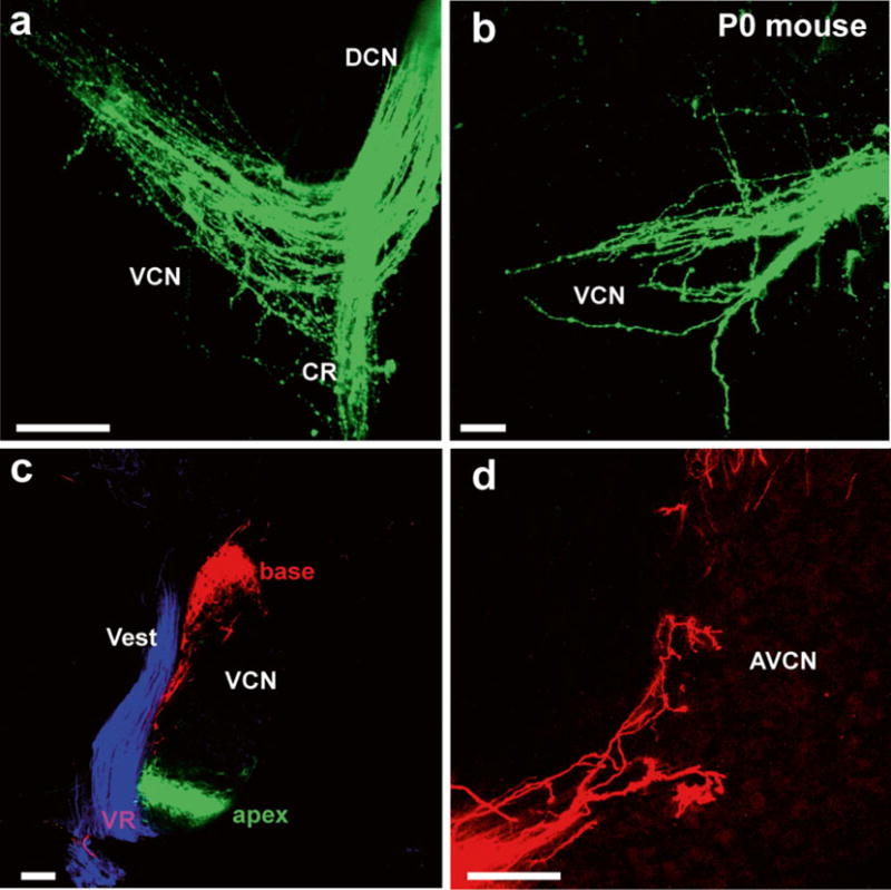
Details of afferents to cochlear nuclei revealed in (a, b) whole mounts and (c, d) sections. Lipophilic dyes can reveal the pattern of innervation of cochlear afferents and deviations from normal projections in mutant mice (a, b) or can be used in vibratome sections (c) to show the segregation of basal, apical, and vestibular projections to the cochlear nucleus and the nearby vestibular nucleus. (d) Higher magnifications show the Golgi-like filling of afferents beginning to form endbulbs of Held. AVCN anteroventral cochlear nucleus, CR cochlear root, VCN ventral cochlear nucleus, Vest vestibular nucleus, VR vestibular root. Bar equals 100 μm. Modified after [67]