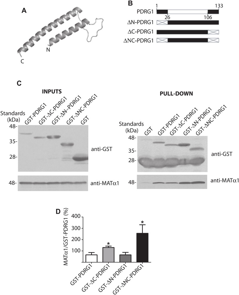
Fig 2. Structural model of rat PDRG1 and interaction of PDRG1 truncated forms with MATα1.
(A) PDRG1 structural model comprising residues K27-Q106 obtained with PHYRE. (B) Schematic representation of PDRG1 and the truncated forms prepared; the modeled area (white box) and deleted sequences (crossed box) are indicated. (C) Representative western blots of pull-down experiments carried out with recombinant truncated PDRG1 forms and MATα1 using anti-GST and anti-MATα1. Incubations with MATα1 were carried out in the presence of excess GST to avoid unspecific binding. The size of the standards is indicated on the left side of the panels. (D) Quantification of the MATα1/GST-PDRG1 signal ratio (mean ± SEM) from seven independent pull-down experiments (*p≤0.05 vs GST-PDRG1).

