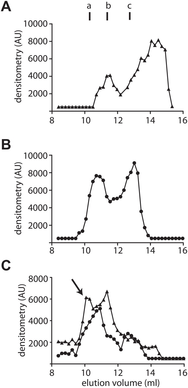
Fig 5. Evaluation of the PDRG1/ MATα1 association in nuclear extracts by analytical gel filtration chromatography.
(A) Elution profile of nuclear extracts overexpressing HA-PDRG1 obtained on a Superose 12 10/300 GL column and analyzed by dot-blot using anti-HA. (B) Elution profile of nuclear FLAG-MATα1 detected using anti-MATα1. (C) Elution profile of nuclear extracts overexpressing HA-PDRG1 and FLAG-MATα1; the arrow indicates the new peak recognized by both antibodies (anti-HA (▲) and anti-MAT (●)). Elution of the protein standards was as follows: blue dextran (7.4 ml); ferritin (9.82 ml); β-amylase (a; 10.62 ml); aldolase (11.1 ml); alcohol dehydrogenase (b; 11.34 ml); conalbumin (c; 12.78 ml); ovalbumin (13.3 ml); carbonic anhydrase (14 ml); lysozyme (17.31 ml); and ATP (17.65 ml). The figure shows representative profiles obtained in five independent experiments.

