Figure 5.

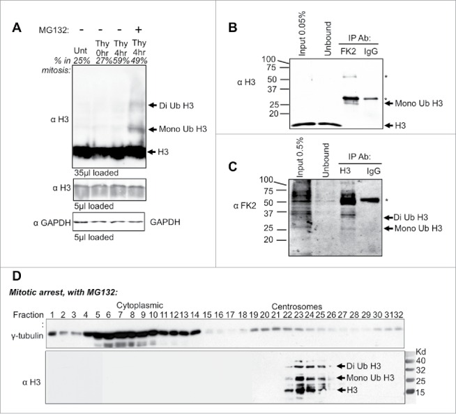
Histone H3 ubiquitin shift detected during mitosis and in purified centrosomes. (A) HeLa cell extracts were harvested at the indicated time points following release from a double thymidine block with or without MG132. Percent in mitosis is indicated above each lane, as determined by flow cytometry analysis. (B) Western analysis of anti-ubiquitin (FK2 antibody) immunoprecipitation from HeLa cells that were double synchronized with Thymidine and released into MG132 for 8 hrs. Immunoblot was probed with histone H3 antibody. Heavy and light chain is marked by “∗.” (C) Western analysis of anti-histone H3 Immunoprecipitation from HeLa cells that were treated as in B. Immunoblots were probed with anti-ubiquitin (FK2 antibody). The heavy chain is marked by “∗.” (D) Velocity sedimentation of HeLa cell extracts upon double thymidine arrested cells released into MG132. Extracts were separated on a 15–60% sucrose gradient.
