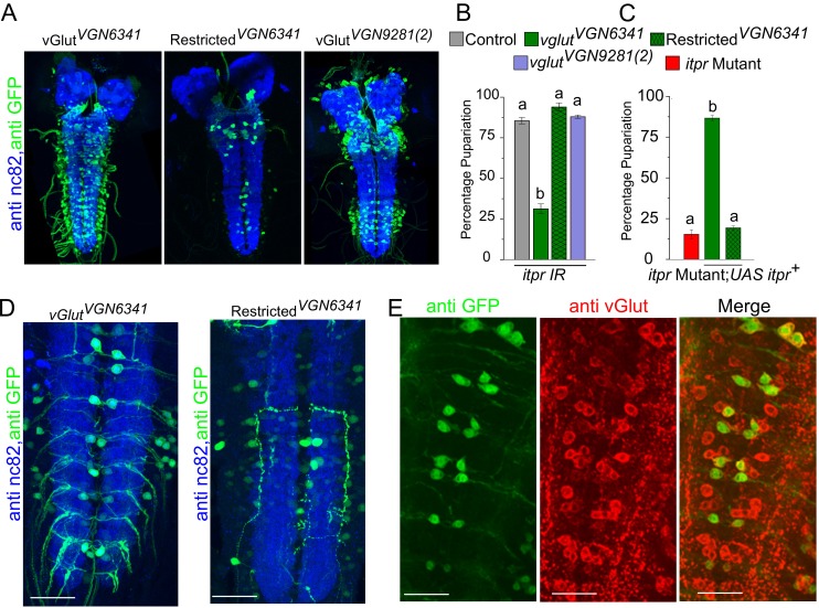
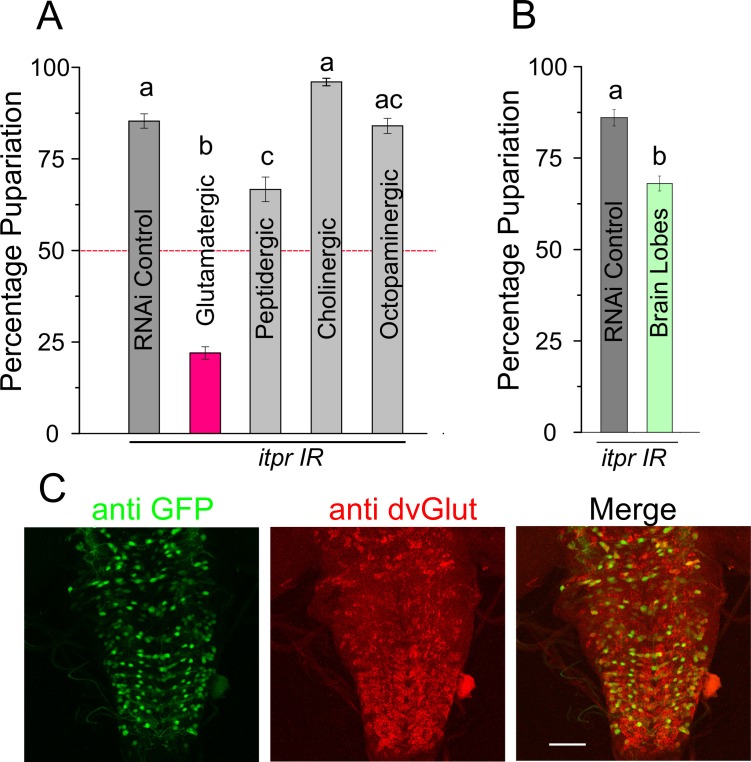
Figure 2. Knockdown of the IP3R in glutamatergic neurons prevents pupariation upon PDD.
(A) Expression patterns of GAL4 drivers used in (B) determined using UAS-eGFP and co-stained with anti-nc82. (B and C) Bars show mean percentage pupariation (± SEM) of the indicated genotypes on PDD. N ≥ 6 batches with 25 larvae each. (D) Images of selected substacks of the ventral ganglion of VGN6341-GAL4, with and without tsh-GAL80, expressing UAS-eGFP, double labelled with anti-nc82. (E) Selected substacks showing overlap of all dvGlut-positive cells and GFP-positive cells marked by VGN6341-GAL4 in the ventral ganglion. Scale bars indicate 50 µm. Bars with the same alphabet represent statistically indistinguishable groups (one-way ANOVA with a post hoc Tukey’s test p<0.05).


