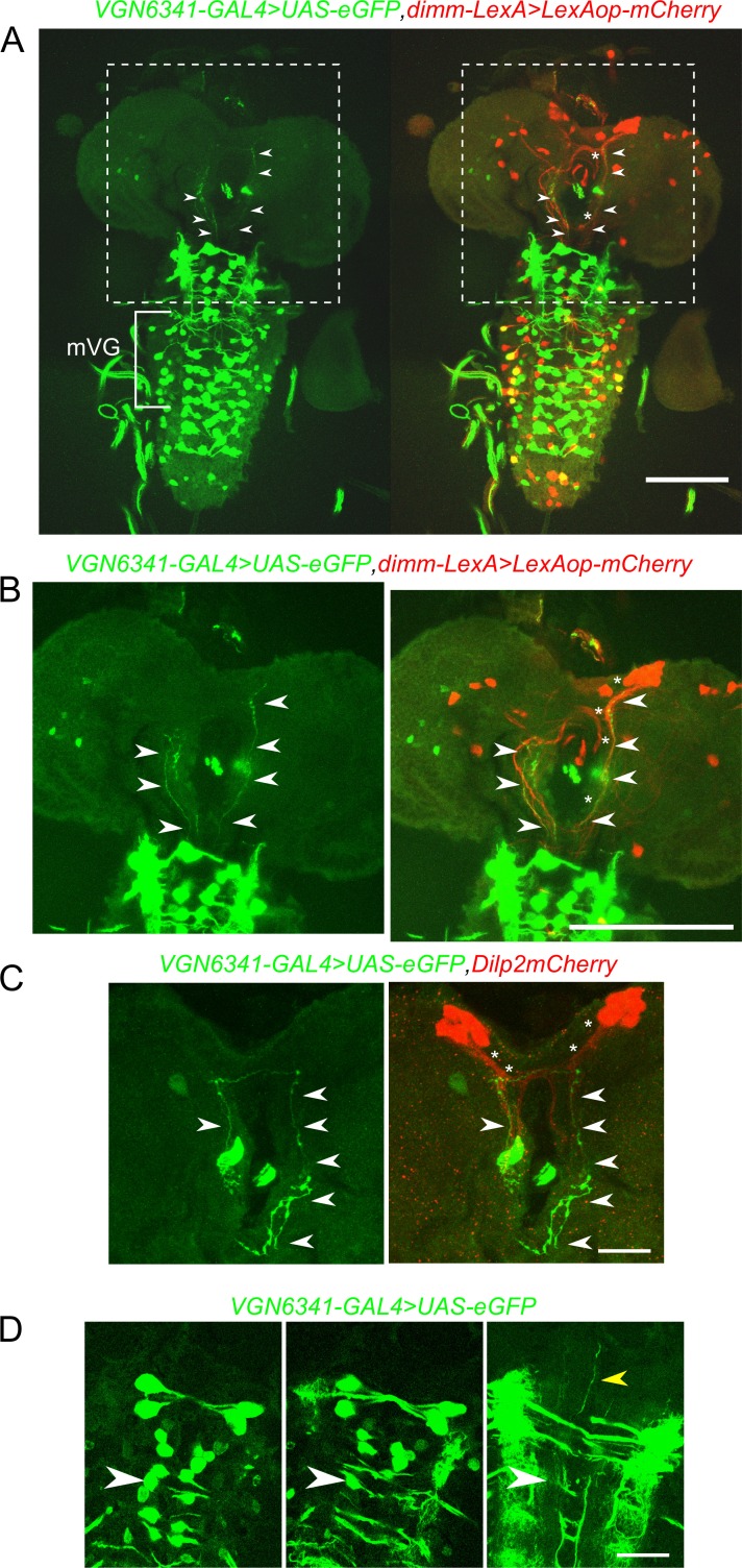
Figure 4. Glutamatergic neurons in the larval ventral ganglion project to peptidergic neurons in the mNSC.
A and B Selected confocal stacks showing the neurites marked by VGN6341-GAL4 driven UAS-eGFP (green) and their merged patterns with dimm-LexA-driven expression of LexAop-mCherry (red). The boxed area in A is shown in B as a high-magnification image. Arrow heads indicate VGN6341-GAL4 expressing neurites projecting toward the mNSCs. Asterisks mark dimm-LexA labelled projections. (C) Neurites marked by VGN6341-GAL4-driven eGFP (arrow heads) overlap with projections of the mNSCs marked by Dilp2mCherry (asterisks). (D) Selected high-magnification confocal images of VGN6341-GAL4 driven UAS-eGFP with an anterior projecting neurite from a midline mVG neuron. The white arrow head marks the same co-ordinates in all three images. The yellow arrow head shows the ascending projections. Scale bars represent 50 µm in A and B and 10 µm in C and D.

