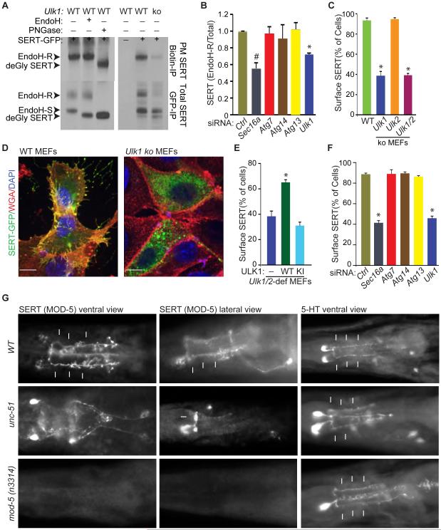
Figure 5. ULKs regulate ER-to-Golgi trafficking.
(A) Representative immunoblots of Biotin IPs (top panels) and GFP IPs (bottom panels) from SERT-GFP—transfected WT and Ulk1-ko MEFs. WT MEFs were incubated in the presence or absence of Endo H or peptide-N-glycosidase F (PNGase) to establish the migration pattern of the different glycosylated forms of SERT. (B) Mean ratios (±SD) of Endo H–R SERT to total SERT in RNAi-treated samples from 2 independent experiments. *P <0.01 and #P <0.05 and (ANOVA) when compared with Ctrl. (C) Mean percentages (±SEM) of cells showing colocalization of AlexaFluor 647–conjugated wheat germ agglutinin (WGA) and SERT-GFP. Data were acquired from 3 independent experiments, and more than 100 cells per population were scored. *P <0.001 (ANOVA) when compared with WT. (D) Representative merged pseudocolored images of SERT-GFP—transfected WT and Ulk1-ko MEFs stained with AlexaFluor 647–conjugated wheat germ agglutinin (WGA) and DAPI. Scale bar: 10 μm. (E) Mean percentages (±SEM) of cells with colocalized WGA and SERT-GFP. Data were acquired from 3 independent experiments, and more than 100 cells per population were scored in each experiment. *P <0.002 (ANOVA) when compared with empty vector–transduced cells. (F) Mean percentages (±SEM) of siRNA transfected cells with colocalized WGA and SERT-GFP. Data were acquired from 3 independent experiments, and more than 100 cells per population were scored. *P <0.001 (ANOVA) when compared with siCtrl-transfected cells. (G) Representative images of WT, unc-51 mutant and mod-5 mutant C. elegans stained with antibodies against MOD-5/SERT and 5-HT. The arrows highlight the 5-HT or MOD-5/SERT staining of NSM processes. See also Figure S5.

