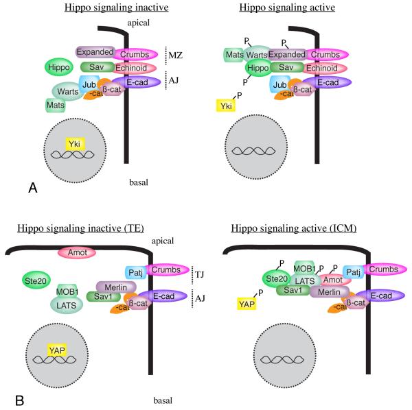
Figure 2. Localization and re-localization of core Hippo pathway components.
A) Localization of core components in Drosophila epithelia. Under conditions of low Hippo pathway activity (left), Wts is associated with its inhibitor, Jub, at adherens junctions (AJ), and Hpo is predominantly cytoplasmic, while Yki is nuclear. Under conditions of high Hippo pathway activity, Wts localizes with Ex at the marginal zone (MZ), and Hpo is recruited to Sav, causing Yki to be cytoplasmic. B) Localization of core components in 32 cell mouse blastocysts. In outer TE cells, Hippo signaling is low, YAP is nuclear, and Amot is localized to the apical membrane. This is presumed to prevent the formation of a LATS activation complex, although the localization of LATS proteins has not been determined in this tissue. In inner ICM cells, Hippo signaling is high, YAP is cytoplasmic, and Motins (Amot) are localized to cell-cell junctions, where, together with Merlin/NF2, they promote phosphorylation and activation of LATS [118].

