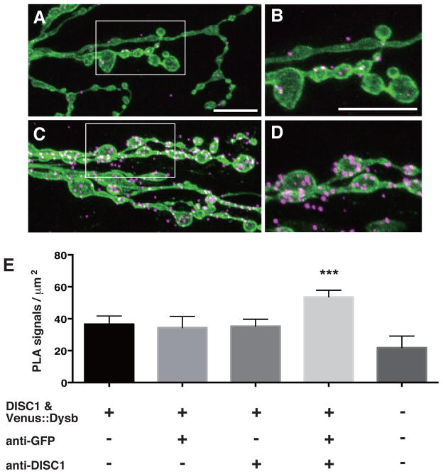
Figure 7. DISC1 directly interacts with the Drosophila Dysbindin in the larval NMJ.
A–D. Confocal images of PLA signals in the larval NMJ. A, B. PLA in w (CS10) control NMJs. C, D. PLA in NMJs expressing Drosophila-Dysbindin and DISC1. Third instar larval NMJ. UAS-Venus::Drosophila-Dysbindin and UAS-DISC1 were co-expressed with tubP-GAL4. B, D. Higher magnification of the area indicated in A and C. Green, motor neuron termini labeled with anti-HRP. Magenta, PLA signals. Scale bars, 10 μm.
E. Quantification of PLA signals. Venus::Dysbindin was detected with anti-GFP. ***p < 0.001 by one-way ANOVA followed by Dunnett’s post hoc test. n = 9–19.

