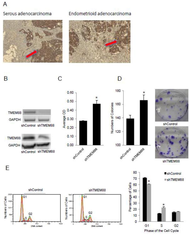
Figure 3. TMEM88 is expressed in ovarian cancer tumors and is associated with cell proliferation.

A. Representative immunostaining for TMEM88 in serous and endometrioid OC (100× magnification). B. RT-PCR measures TMEM88 mRNA expression level in SKOV3 cells stably transduced with shRNA targeting TMEM88 (shTMEM88) vs control shRNA (shControl). Western blot measures TMEM88 protein level expression in shTMEM88 and shControl transduced SKOV3 cells. C. Proliferation of SKOV3 cells stably transduced with shTMEM88 or shControl. D. Numbers of colonies formed by SKOV3 cells stably transduced with shTMEM88 or shControl. Bars represent means ± SE. * denotes statistical significance (p < 0.05). E. Cell cycle analysis by flow cytometry in SKOV3 cells transduced with shControl and shTMEM88. Bars represent means ± SD, * denotes statistical difference (p < 0.01).
