Figure 2.
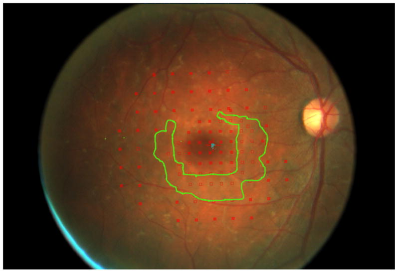
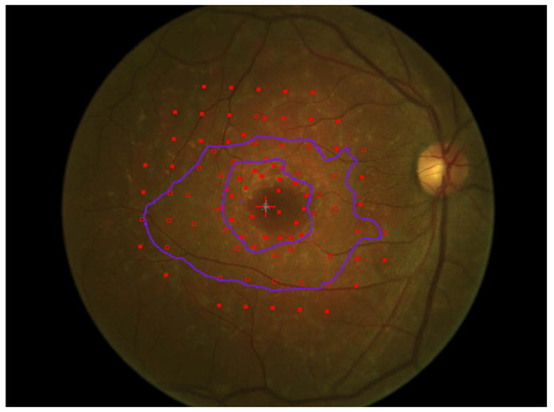
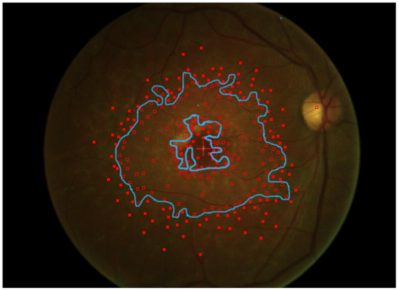
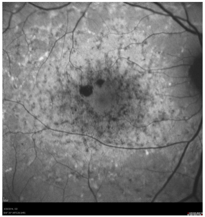
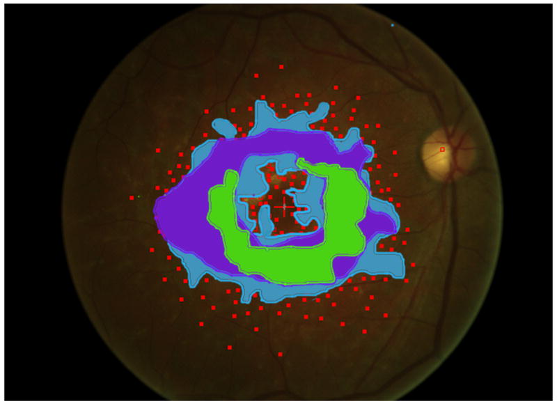
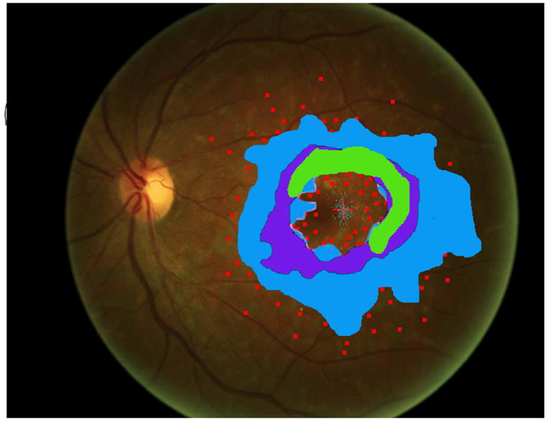
Mapping the dense scotoma. This 50 year old patient (patient 7 on the table and other figures) had a partial macular ring scotoma at baseline (a), which evolved into a ring scotoma 3 years later (b). At the last visit, 8 years after baseline, (c), there is a larger macular ring scotoma with preservation of the foveal region. Fundus autofluorescence done at the time of the last visit (d) shows only two small homogeneously dark areas. The rest of the image, although abnormal, does not provide information as to where scotomas might be. Figures e and f show the dense scotoma as solid filled areas colored based on the year tested for the right eye and left eye respectively. There is still sparing of both foveas, with greater sparing in the left eye.
