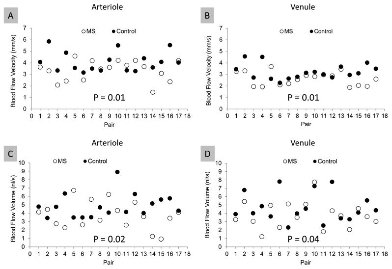
Figure 2. Scatterplot of retinal blood flow velocity and perifoveal flow volume of MS patients compared with age- and gender-matched controls.

One eye of each subject was imaged with the RFI device, and blood flow velocities in arterioles and venules in the perifoveal region were measured. The blood flow volume of the perifoveal region was also calculated based on the vessel diameter crossing a circle of 2.3-mm, centered at the fovea. The mean blood flow velocities in MS subjects were significantly lower than controls in both the arterioles (A) and venules (B). Similarly, the mean perifoveal blood flow volume of both arterioles (C) and venules (D) were significantly lower in MS subjects than in controls. In both groups, the blood flow velocity was higher in arterioles than venules (A and B, P = 0.001). In contrast, the perifoveal blood flow volume of the arterioles was similar to that in the venules in both groups (C and D, P = 0.36).
