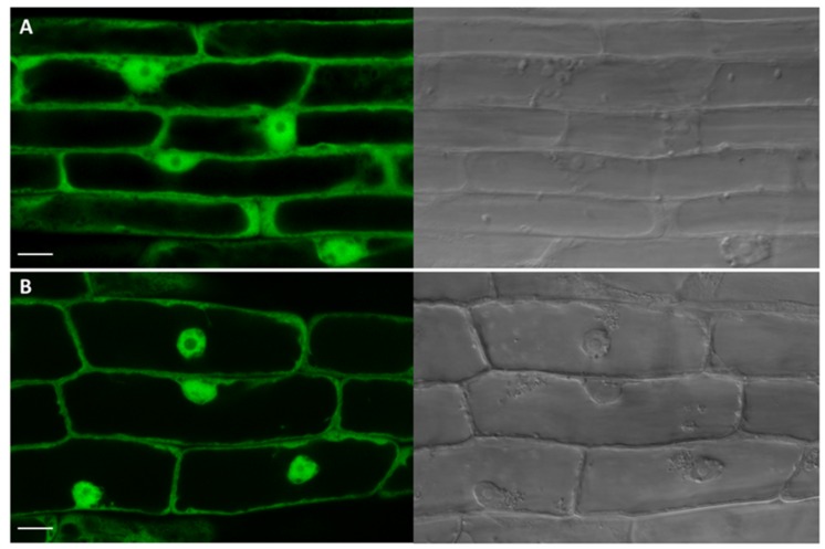
FIGURE 9.
Subcellular localization analysis of UGT76C2-GFP fusion protein. Green channel and transmission light images captured by confocal microscopy. (A) A control root overexpressing SU:GFP with typical GFP pattern in cytosol and nuclei. (B) The root cells overexpressing SU:UGT76C2-GFP with signal indicating the cytosolic localization of UGT76C2. Scale bars 10 μm.

