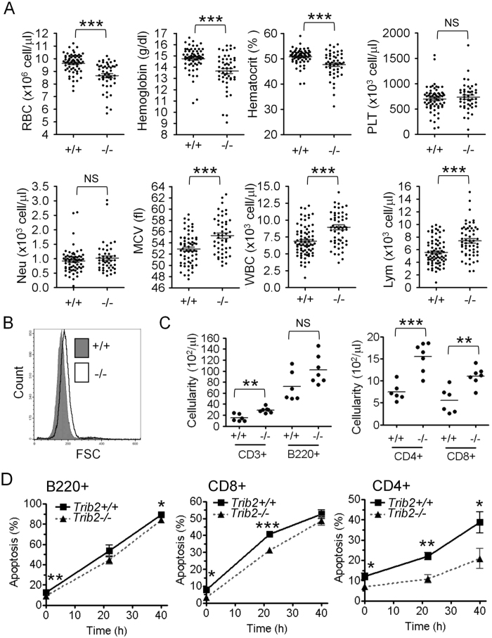
Figure 1. Trib2 deficiency confers macrocytic anemia on mice.
(A) Complete blood count analyses of peripheral blood from Trib2+/+ (n = 63) and Trib2−/− (n = 49) mice. 260 μL of blood was withdrawn into sodium citrate-containing tubes and subjected to complete blood counts using Abbott Cell-Dyn 3700. (B) Analysis of RBC cell volume of Trib2+/+ and Trib2−/− mice by flow cytometry. 1 μL of blood was diluted with 200 μL of serum-PBS solution and analyzed by flow cytometry. (C) Numbers of CD3+, B220+, CD4+ and CD8+ cells in peripheral blood of Trib2+/+ (n = 6) and Trib2−/− (n = 7) mice. WBCs were isolated from peripheral blood and characterized for specific surface markers by flow cytometry, as indicated. Absolute numbers of cells were calculated by multiplying the relative proportion of a particular cell population with the absolute number of WBCs. (D) Cytokine withdrawal-induced apoptosis in peripheral lymphocytes. WBCs were isolated from peripheral blood of Trib2+/+ and Trib2−/− mice and cultured in cytokine-free medium in vitro. Apoptosis of B220+, CD8+, and CD4+ cells were analyzed by Annexin-V staining and specific surface markers by flow cytometry (n = 8 for each genotype). RBC, red blood cell; PLT, platelet; Neu, neutrophil; MCV, mean corpuscular volume; WBC, white blood cell; Lym, lymphocyte. The graphs show the mean and S.E.M., *P < 0.05 compares between the indicated groups. **P < 0.01 compares between the indicated groups. ***P < 0.005 compares between the indicated groups. NS: no significant difference.

