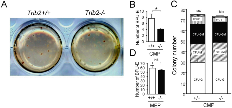
Figure 5. Deletion of Trib2 decreases BFU-E colonies of CMPs, but not MEPs.
(A–C) CMPs were purified from Trib2+/+ and Trib2−/− mice as described in Fig. 3A and subjected to an in vitro colony-forming unit (CFU) assay for differentiation into myeloid and erythroid lineages. (A) Representative images of CFU plates are shown. The sorted Trib2+/+ or Trib2−/− CMPs were cultured in methylcellulose-based medium with recombinant cytokines and erythropoietin for 10 days. (B) The numbers of BFU-E derived from 200 sorted Trib2+/+ and Trib2−/− CMPs were recorded from plates in (A) (n = 6). (C) Bar graphs show the number of each different type of colony observed in plates (A). (D) 200 sorted MEPs from Trib2+/+ and Trib2−/− mice were cultured in vitro as described in (A), and the number of BFU-E was recorded (n = 4). The graphs show the mean and S.E.M. *P < 0.05 compares between the indicated groups. NS: no significant difference.

