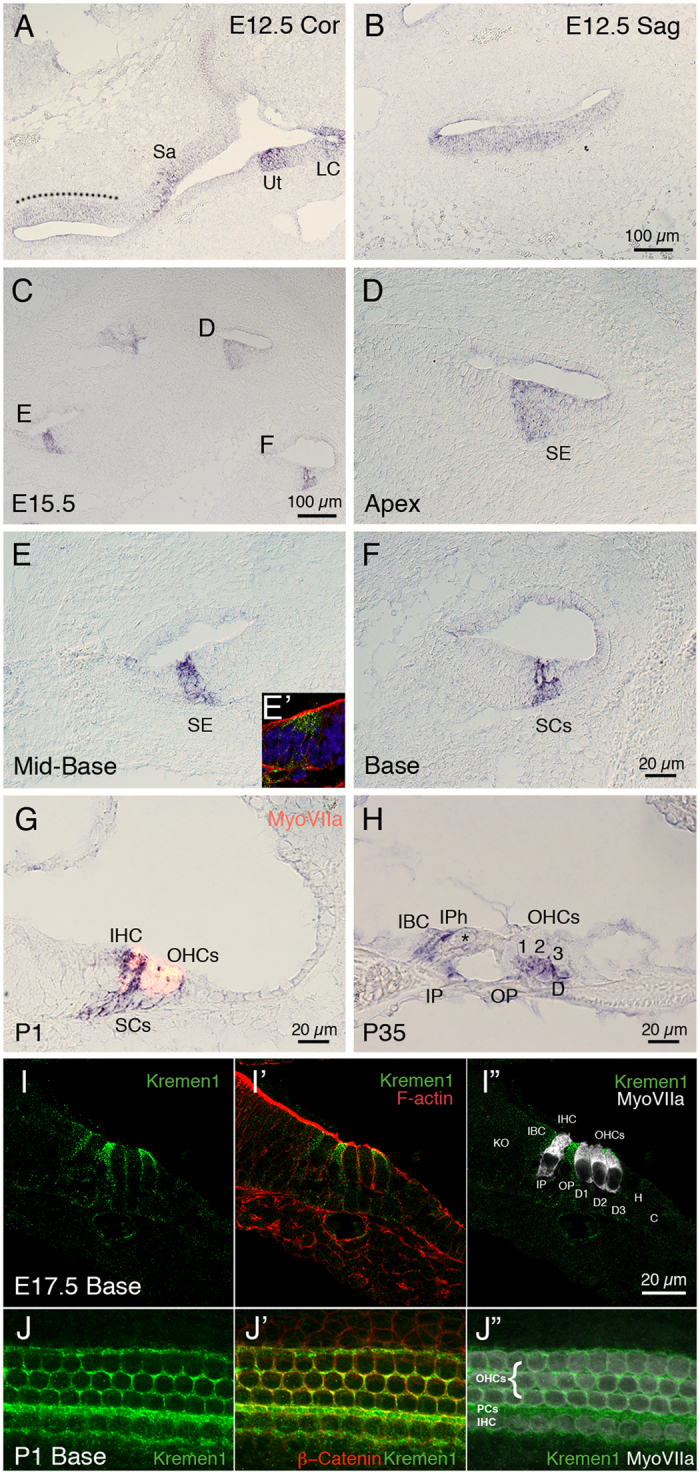
Figure 1. Kremen1 is expressed in the developing and adult mammalian cochlea.

(A) Coronal section through the head at E12.5. Kremen1 expression is detected in the floor of the cochlear duct (dotted line) and prosensory region of the vestibular system. (B) A sagittal section through the head at E12.5, Kremen1 expression is found in the floor of the cochlear duct. (C) A low magnification view of a transverse section through an E15.5 cochlea showing Kremen1 expression in all turns. (D) Kremen1 expression in the prosensory region of the apical turn of an E15.5 cochlea. (E) Kremen1 expression in the prosensory region of the midbasal turn of an E15.5 cochlea. (E’) Immunohistochemistry for Kremen1 (green), cell membrane (actin, red) and cell nuclei (Dapi, blue) in the midbasal turn on E15.5. Kremen1 protein localized to the prosensory region. (F) Kremen1 expression in the supporting cell region of the base of an E15.5 cochlea (D, E, scale as in panel F). (G) High magnification view of transverse sections through P1 cochlea showing Kremen1 expression in supporting cells. Hair cells are counterstained for MyoVIIa, in red. (H) High magnification view of transverse section through P35 cochlea. (I,I’,I”) High magnification view of transverse section through E17.5 cochlea showing immunohistochemistry for Kremen1 (green), cell membrane (actin, red and hair cells (MyoVIIa, white). (J,J’,J”) High magnification view of the luminal surface of the basal region of a P1 cochlea showing immunohistochemistry for Kremen1 (green), cell membrane (β-Catenin, red) and hair cells (MyoVIIa, white). SE; sensory epithelium, Sa; saccule, Ut; utricle, LC; lateral canal, IHC; inner hair cell, OHC; outer hair cell, IBC; inner border cell, IP; inner pillar cell, IPh; inner phalangeal cell, OP; outer pillar cell, PCs; Pillar cells, D; Deiters’ cells, SC; supporting cell.
