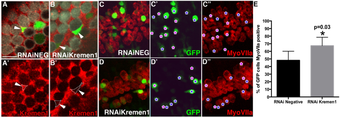
Figure 3. Loss of Kremen1 made a prosensory cell more likely to develop as a hair cell.
(A,B) High magnification surface view of negative control RNAi construct (A) or Kremen1RNAi (B) transfected cochlear explant, hair cells are labeled by antiMyoVIIa (white), supporting cells are labeled by antiKremen1 (red), transfected cells express GFP (green). (A) Arrowhead indicates a supporting cell transfected with RNAiNeg construct that expresses Kremen1 on its surface at similar levels to non transfected neighboring cells. (A’) Single channel view of (A). (B) Arrowheads indicate supporting cells transfected with Kremen1RNAi that display very little Kremen1 protein staining relative to their non-transfected neighbors. (B’) Single channel view of (B). Scale bar indicates 10 μm. (C,D) Surface view of apical region of a cochlear explant electroporated on E12.5 and cultured in vitro for six days, with either negative control RNAi (C,C’,C”) or Kremen1RNAi (D,D’,D”). Hair cells are labeled by antiMyoVIIa (red), transfected cells express GFP. Purple asterisks (*) show GFP cells that do not express MyoVIIa, blue asterisks mark GFP positive cells that express MyoVIIa. (E) Quantification of transfected cells expressing MyoVIIa. Asterisk denotes p < 0.05. All error bars represent standard deviation.

