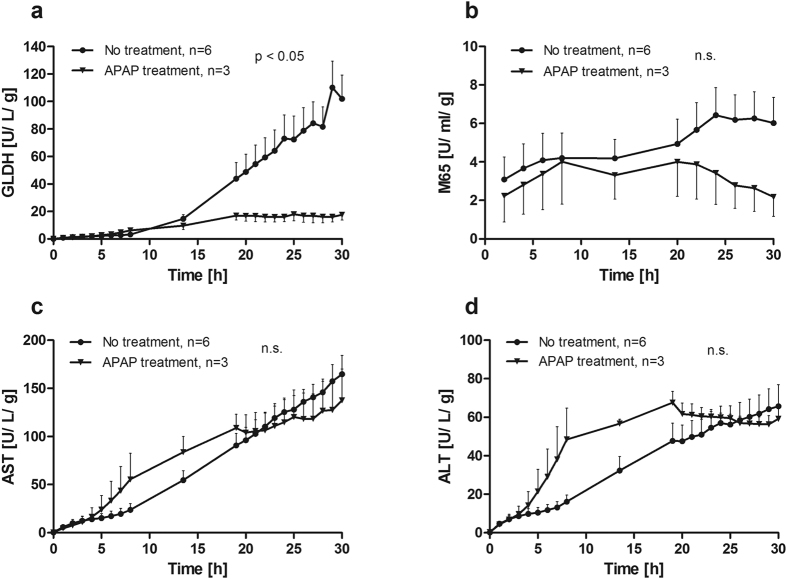
Figure 4. Course of markers for liver cell damage during perfusion.
Release of (a) glutamate dehydrogenase (GLDH), (b) cytokeratin-18 (M65), (c) aspartate aminotransferase (AST) and (d) alanine aminotransferase (ALT) into the perfusate were detected. More GLDH and M65 were released from untreated liver specimen compared to acetaminophen treated (APAP) livers while AST and ALT did not differ between the two groups (two-way ANOVA).

