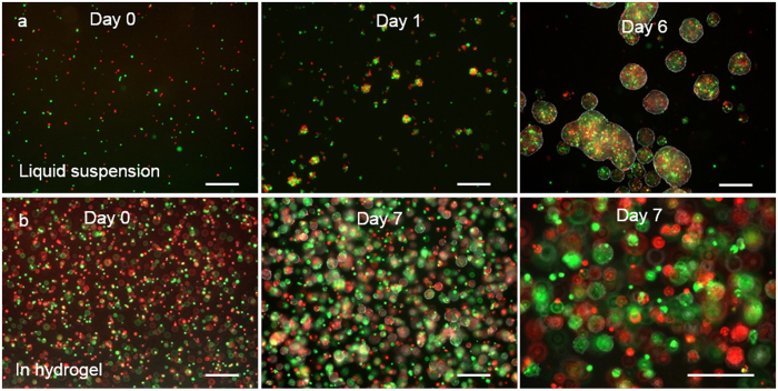
Figure 2.
Culture glioblastoma TICs in 3D. L0 TICs stained with green or red fluorescent dye were mixed at 1:1 and cultured in suspension in liquid medium statically at 5 × 10⁴ cells/ml (a) or in the thermorevesible PNIPAAm-PEG hydrogel at 1 × 10⁶ cells/ml (b). Spheroids in the suspension culture contained both green and red cells (a) while spheroids in the hydrogel contained either green or red cells (b). Scale bar: 250 μm.

