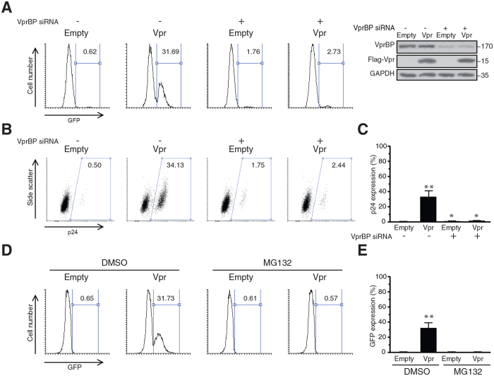
Figure 3. Vpr-induced reactivation of HIV-1 is VprBP and proteasome-dependent.
(A–C) J-Lat 10.6 cells were transfected with siRNA against VprBP or non-targeting siRNA. After 48 h, cells were transduced with lentiviral vectors for expression of Vpr or empty vectors. Twenty four hours post-transduction, cells were analyzed for expression of GFP. Some cells were also spared and analysed using Western blot for depletion of VprBP (A). The intracellular p24 was labeled 48 h post-transduction and cells were analysed using flow cytometry (B). (C) Mean of 3 independent experiments as described in (B). (D) J-Lat 10.6 cells were transduced with lentiviral vectors for expression of Vpr. Transduced cells were treated with 5 μM MG132 or DMSO. After 24 h, HIV-1 reactivation was analysed by measuring the expression of GFP. (E) Mean of 3 independent experiments as described in (D).

