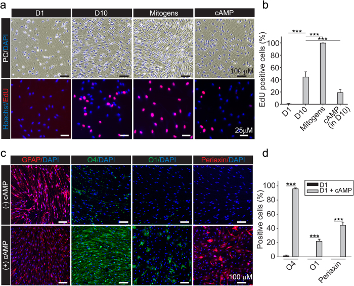
Figure 7. Analysis of proliferation and differentiation of isolated adult SCs obtained after cryopreservation.
(a,b) Determination of the mitogenic responses to serum and soluble growth factors. Cells were plated as described in Fig. 6 in DMEM containing a non-mitogenic concentration (1%) of FBS (D1), 10% FBS (D10), 10% FBS supplemented with neuregulin and forskolin (Mitogens) or D10 in the presence of 250 μM CPT-cAMP (cAMP). Cells were monitored by phase contrast microscopy (upper panels) to reveal changes in cell morphology, alignment and density in each experimental condition. Treatment was carried out for 3 days. Results from EdU incorporation assays are shown in (a) (lower panels) and (b) (quantification of fluorescence microscopy data). (c,d) Determination of the differentiating responses to prolonged treatment with cell permeable cAMP analogs. Cells were plated as described in (a,b) with the exception of using D1 medium containing vehicle (control, −cAMP) or CPT-cAMP (250 μM) as inducer of differentiation (+cAMP). Cells were analyzed for the expression of the indicated markers by immunofluorescence microscopy 4 days after stimulation (bars = mean ± SD, ***p < 0.001).

