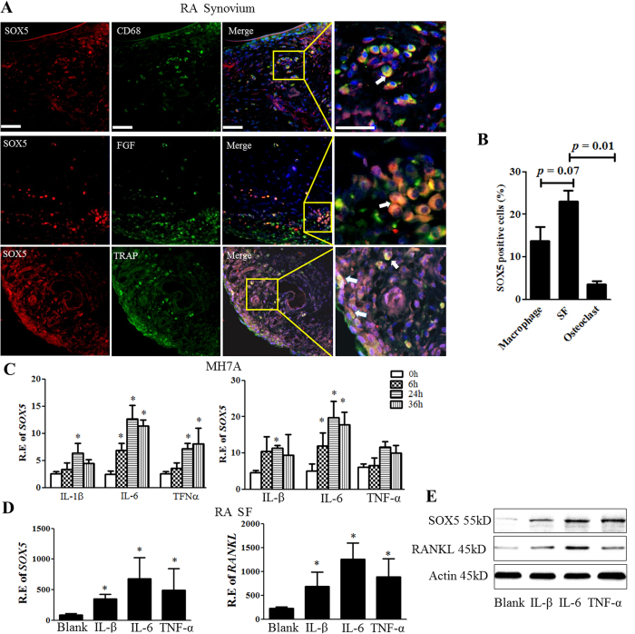
Figure 2. SOX5 is up-regulated by pro-inflammatory cytokines in SF of RA synovium.
(A) Representative immunofluorescence microscopy images of SOX5 and macrophage marker CD68 (upper), SF marker FGF (middle) or osteoclast marker TRAP (lower) in RA synovium. Scale bar, 100 μm. The arrows point at double-stained cells. (B) The percentage of macrophage, SF and osteoclast positive for SOX5staining. (C) MH7A cells were treated with IL-1β (100 ng/ml), IL6 (100 ng/ml), or TNFa (100 ng/ml) for 0–36 h. SOX5 (left) and RANKL (right) expression were quantified by Real-time PCR (n = 3). (D) Primary cultured SF were treated IL-1β (100 ng/ml), IL6 (100 ng/ml), or TNFa (100 ng/ml) for 24 h, and SOX5 (left) and RANKL (right) mRNA levels were quantified (n = 6). Values are means ± SD (*p < 0.05). (E) Representative images of western-blot detection of SOX5 and RANKL protein expression of in MH7A after IL-1β, IL6, or TNFa treatment for 24 h. All experiments were performed in triplicate and were repeated three times.

