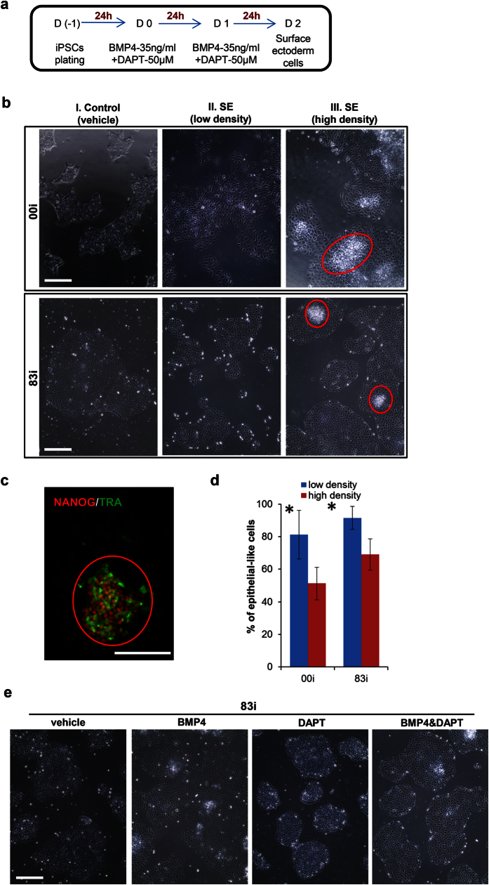
Figure 1. Induction of surface ectoderm (SE) differentiation from hiPSCs.
(a) Protocol for in vitro differentiation of hiPSCs to SE. (b) Cell morphological changes after SE induction. The top and bottom panels show the morphological changes in undifferentiated and differentiated hiPSC lines 00i and83i, respectively. Red circles: undifferentiated hiPSCs. Original magnification x100. (c) Representative immunofluorescence image shows undifferentiating cells under high density condition expressing pluripotent markers NANOG and TRA-1-81. Original magnification x200. (d) Percentile of epithelial-like (differentiated) after 48 h SE induction in 00i and 83i hiPSCs. hiPSCs were induced to SE differentiation in low or high density. After 48 h induction, number of epithelial-like and iPSCs-like morphologies was counted under high magnification (x200) from five random fields. % of epithelial-like cells was calculated as following: % = (number of epithelial-like cells/total number of cells) x100 and plotted as (mean ± SD). *p < 0.05. (e) Representative images displayed cell morphologies treated by vehicle (DMSO), BMP4, DAPT or (BMP4 + DAPT) after 48 h. Original magnification x100. Bars: 100 μm.

