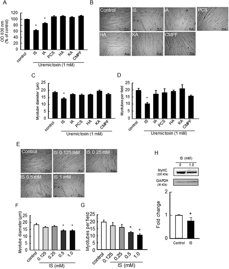
Figure 1. Effect of uremic toxins on the proliferation and myotube formation of C2C12 myoblast cells.
(A) Effect of uremic toxins on C2C12 myoblast cell proliferation was determined by a methylene blue assay. C2C12 myoblast cells were seeded on a 96-well plate and cultured overnight. After adhesion, cells were treated with uremic toxins (1 mM) and incubated for 72 hr. Adherent cells were fixed and stained with methylene blue. The OD630 nm was then measured. Effect of uremic toxins on cell differentiation was determined by C2C12 myoblast cell tubular formation. C2C12 myoblast cells were seeded on a 12-well plate and cultured overnight. After cell adhesion, cultured medium was changed to differentiation medium containing with uremic toxins and cultured for 7 days. The cell density in each treatment group was the same before changing the culture medium to differentiation medium. (B) Cell morphology was observed by microscopy, and (C) average diameters of at least 50 myotubes were determined to be 40 μm along the length of the myotube. (D) The number of the myotubes per field was measured 5 fields per sample. Myotubes were counted, and diameter was assessed on Image J. Dose-dependent effect of IS on (E) cell morphology, (F) myotube diameter, and (G) the number of myotubes were shown. Scale bar = 100 μm. (H) Myotube formation was also evaluated using second marker, namely, the myosin heavy chain (MyHC) by Western blot. Data are expressed the means ± SEM (n = 3~6). *p < 0.05, **p < 0.01 compared with control.

