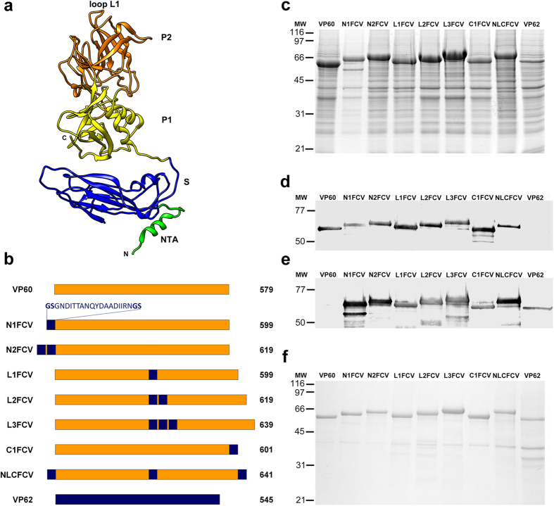
Figure 1. Expression and characterization of VP60 insertion mutants harbouring a FCV capsid protein B-cell epitope.
(a) Ribbon representation of the VP60 protein structure (Protein Data Bank [PDB] accession number 3J1P). The NTA, S domain, P1 and P2 subdomains, and loop L1 are indicated. (b) Schematic representation showing names (left) and protein lengths in amino acids (right). The amino acid sequence depicted (FCV B-cell epitope) was inserted at the indicated positions in each VP60 insertion mutant. RHDV capsid protein (VP60) and FCV capsid protein (VP62) are also shown. (c) H5 cells were infected with each recombinant baculovirus and infected-cell lysates were analyzed by SDS-10% PAGE. (d,e) Western blots performed using a rabbit hyperimmune serum against RHDV to detect VP60 protein (d), or a monoclonal antibody directed against the FCV B-cell epitope (e). (f) Infected H5 cell cultures were subjected to VLP-purification procedures and the resulting samples were characterized by SDS-10% PAGE. Molecular weight markers (MW; ×103 Da) are given on the left.

