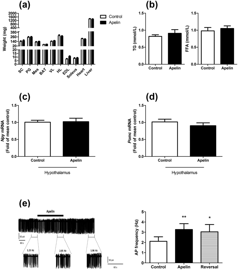
Figure 2. Apelin depolarizes POMC neurons in the hypothalamus without effect on food intake and tissue weight.
Effect of chronic apelin treatment (Apelin) versus chronic aCSF treatment (Control) (a) on tissues weight (SC: subcutaneous adipose tissue, PG: perigonadal adipose tissue, Mes: mesenteric adipose tissue, BAT: brown adipose tissue, VL: vastus lateralis (muscle); HL: muscle; EDL: extensor digitorum longus (muscle)); (b) on triglycerides (TG) and free fatty acids (FFA) plasma levels; (c) on hypothalamic Npy mRNA expression and; (d) on hypothalamic Pomc mRNA expression. (e) (Left panel) Representative cell-attached recording of a POMC-GFP neuron activated by apelin (thick black bar under the trace). Panels below the trace represent enlarged 60 seconds recording periods with average action potential frequency before, during and after apelin bath application. (Right panel) Quantification of action potential (AP) frequency of POMC before (control; over the last 60 s before apelin application), during (apelin, over the last 60 seconds of apelin application) and after (reversal, over 60 seconds, 5 minutes after apelin application) apelin application. Experiments were performed with a set of 6–9 mice in each group. **p < 0.01; *p < 0.05 vs. control.

