Figure 1.
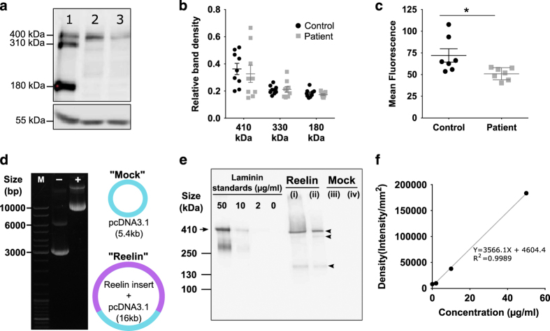
Patient-derived cells have reduced endogenous reelin and recombinant full-length reelin production into conditioned medium. (a) Qualitative representation of reelin expression by western blot showing total protein samples obtained from (1) mouse brain lysate, (2) healthy control olfactory neurosphere-derived (ONS) cell line and (3) schizophrenia patient ONS cell line. Mouse brain lysate was used as a positive control and β-tubulin (55 kDa) was the loading control. (b) Quantitation of reelin expression by western blot. Reelin band densities were divided with β-tubulin band densities to obtain relative band intensities. Relative band intensities were presented as mean±s.e.m. (c) Flow cytometry quantitation of reelin expression in fixed ONS cells. Fluorescence intensities for 10,000 cells stained with anti-reelin antibody followed by Alexa Fluor 488 were measured via flow cytometer and normalized to isotype-matched IgG antibody. (d) Amplification and purification of full-length reelin plasmid pCrl and mock vector pcDNA3.1. Plasmids were verified by ethidium bromide agarose gel. GeneRuler DNA ladder mix was used as the size marker (“M”); other two lanes contained “Mock” vector pcDNA3.1 (−) and “Reelin” plasmid pCrl (+). (e) Verification of recombinant full-length reelin. Conditioned medium was collected from HEK293FT cells transfected with either the “Reelin” pCrl plasmid or “Mock” plasmid. Recombinant mouse laminin (molecular size 410 kDa; arrow) was used as a standard to validate full-length reelin size and to estimate concentrations present. Conditioned medium was purified and concentrated with Amicon-15 centrifugal tubes. Unpurified samples presented in lanes (i) and (iii) for reelin and mock-conditioned medium, respectively. Purified reelin and mock-conditioned medium were in lanes (ii) and (iv). Reelin bands were probed with anti-reelin antibody to give the full-length band (410 kDa) and its isoforms, highlighted with arrowheads. (f) Laminin standard curve to correlate western blot band density (y axis) with protein concentration (x axis). All data were presented as mean per group±s.e.m. Each point in the scatter plot represents data for each cell line used. *Student’s t-test, P=0.028.

